Now available at Breach Candy Hospital on Wednesdays 07:00 pm to 08:00 pm

Dr. Dilip Raja
Urologist & Andrologist
Ureteric Stones
In India, approximately 5 -7 million patients suffer from stone disease
Get in touch with us today (022) 2645 2007
URETERIC STONE
The ureter is a thick-walled narrow cylindrical tube which connects the kidney with the urinary bladder. The ureter is approximately 25 to 30 cm in length with 3 – 4mm diameter. It brings down the urine from the kidney into the bladder. The ureter is narrow at 3 junctions and urines passes through it in peristaltic waves. The backflow of urine into the kidney is prevented by ureterovesical valves.
Symptoms And Signs
- Incidental diagnosis on routine health check-ups.
- Dull aching pain in the back.
- Acute colic – This colic pain begins from the flank or the side of mid-back and comes forwards to the groin (from loin to groin). This pain is considered to be worse than labour pain, accompanied by nausea, vomiting and gaseous distension.
- Urinary tract infection.
- Increased frequency of urine.
- Pain and or burning while passing urine.
- Passage of blood in urine (Haematuria).
Investigations
- CBC, Renal Profile
- Routine urine analysis
- Crystals in urine
- Blood Cells
- Puss cells in urine
- Urine for Culture Sensitivity – To rule out UTI and select best antibiotics to treat the infection.
- Ultrasonography of Kidney, Ureter and Bladder – To show the size and swelling (Hydronephrosis) of the kidney and the ureter in obstructive uropathy. It does not give information of function of kidney.
- Intravenous Urography (IVU) – To detect the size and site of stone along with function of kidney. This is the specialized test were series of X- ray are taken after injecting the special medicine, a dye – Contrast, which has radiopaque property. The kidneys excrete these contrasts and kidneys are outlined on X rays and serial films are taken. This is a very useful test. It gives lot of information including size and shape of kidneys, function of kidney – comparative and individual, presence of obstructive uropathy, delineates the anatomy of kidney, ureter & bladder etc.
- CT Scan of Abdomen Pelvis /CT Urography – with or without oral or intravenous contrast. It also gives density of the renal stones in Hounsfield units ( HU). It also gives the function of the kidney. CT scan without contrast has some limitations.
Treatment Of Ureteric Stones
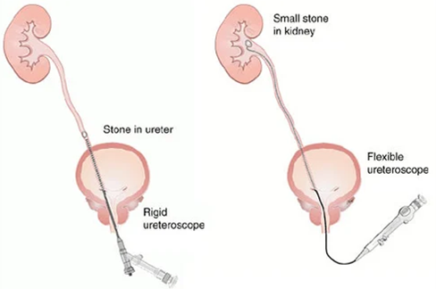
Ureterorenoscope (URS)
Ureteroscopy is highly successful procedure for the retrieval of stone in the ureter. This is a routine procedure performed by urologists. It is passed through the normal urinary opening through the bladder into the ureter. The most common indication is to treat ureteric calculi specially the once which cannot be treated by ESWL or conservative treatment.
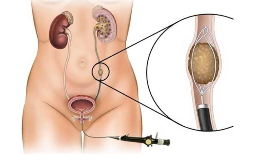
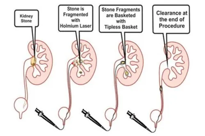
Ureterorenoscopy (URS) involves the passage of an instrument namely Ureteroscope through normal urinary passage. The Ureterorenoscope is advanced under vision through the normal urinary passage under anaesthesia. The Ureterorenoscope is advanced on the side of the ureteric stone and up to the ureteric stone. Once the ureteric stone is localized, various options are available. If the ureteric stone is small, it can be picked up by the forceps & pulled out. But, if the ureteric stone is larger, the ureteric stone can be broken into tiny fragments using Laser technology. Double J stent is usually kept post procedure to drain the kidney. It is a very safe procedure in experienced hands and Ureterorenscopy can treat almost all the ureteric stones.
URS is often used as a diagnostic tool for stones as well as a diagnostic tool for ureteric cancer (tumour). It is a minimally invasive method of treating kidney and ureteric stone.
Ureteroscopy are of two types:
- Rigid Ureteroscopy
- Flexible Ureteroscopy
Retrograde Intrarenal Ureteroscopic Surgery (RIRS)
Flexible Ureterorenoscopy is now available and this flexible ureteroscope can be passed through the normal ureter opening all the way up on the kidneys and stones in the calyces of the kidney can be fragmented into fine particles using laser technology. This procedure required general anaesthesia, spinal or epidural anaesthesia. Hospitalization is generally for 1 or 2 days only.
Open Surgery
Open Surgery for Ureteric Stone are extremely rare nowadays. However, they may be required in large ureteric calculi not amenable to any Endoscopic procedure.