Best Kidney Stone Specialist In Mumbai | Advanced Kidney Stone Treatment – Dr. Dilip Raja
Dilip Raja, a leading kidney stone specialist in Mumbai, provides advanced treatment for kidney stones, including laser stone removal, shockwave lithotripsy (ESWL), and minimally invasive surgery. With years of expertise in urology, he ensures precise diagnosis and personalized care to help patients manage and eliminate kidney stones effectively. Whether you’re experiencing severe pain or recurring stones, Dr. Raja offers the latest medical solutions for quick relief.
Safe and Effective Kidney Stone Treatment with Dr.Dilip Raja Best kidney stone specialist in Mumbai | Urologist. Advanced laser, Surgical & Non Surgical treatments. Schedule An Appointment Today!
Get in touch with us today +91 9820074649
Kidney stones constitute one of the commonest diseases in our country and pain due to kidney stones is known as worse than that of labor pain. In India, approximately 5 -7 million patients suffer from stone disease and at least 1/1000 of Indian population needs hospitalization due to kidney stones.
Geographical Distribution
- The kidney stone disease is widespread particularly in countries with dry, hot climate. These “stone belt regions” of the world are located in countries of Middle East – Dubai, Sharjah, Qatar, Muscat, Abu Dhabi, Saudi Arabia, North Africa, the Mediterranean Regions, North Western state of India and Southern State of USA and areas around the great lakes.
- In India, the “stone belt” occupies parts of Maharashtra, Gujarat, Punjab, Haryana, Delhi and Rajasthan. In these regions, the disease is so prevalent that most of the members of a family will suffer from kidney stones sometime in their lives. Removal of Kidney, Ureteric and Bladder stone procedure forms one of the commonest procedures in Urology department of the hospitals in these regions.
Causes Kidney Stone
Apparently, there are no direct causes for stone formation. However, there are few hypothesis in this regard.
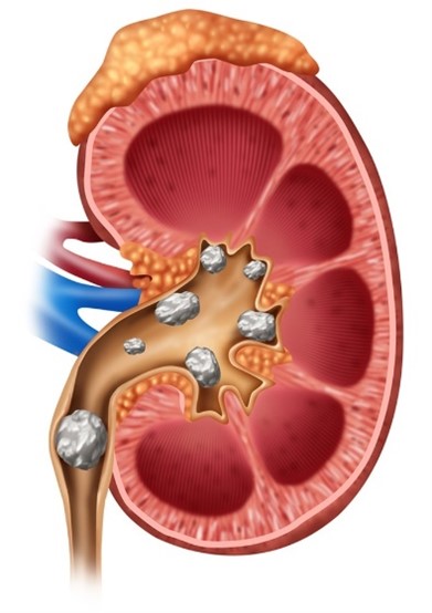
- Kidney Stones: Stones can form anywhere in the urinary system, including the kidneys, ureters, and bladder. They form through a process called supersaturation of urine.
- Hereditery :This may have some role as some people in the same family are more prone to form kidney stones.
- Diet : Diet is not a dominating factor. However, if an individual is a stone former the diet rich in calcium, oxalate & uric acid may increase the chances of stone fornation. In normal individual, diet will not play much role. More than the diet; water intake may be more responsible for kidney stone formation.
- Water Intake: If an individual is a stone former then an increased intake of water will help him pass small gravels before they become nidus for stone formation. Unfortunately, in stone belt region due to dry climate the water is hard and will, in fact, contribute to the formation of stones, if taken in large quantities.
- Medications: Medications like diuretic, excess calcium containing antacids or calcium pills will increase the chances of forming stone formation.
- Other chronic medical illness: Some chronic illnesses are associated with kidney stone formation; in particular, conditions such as cystic fibrosis, renal tubular acidosis, and inflammatory bowel diseases increase the risk of kidney stone formation.
Types Of Kidney Stones
There are various types of urinary stones, but the most common ones are
- Calcium oxalate.
- Uric acid.
- Struvite.
- Cystine stones.
Symptoms And Signs
- Incidental diagnosis on routine health check-ups.
- Dull aching pain in the back.
- Acute colic begins as pain in the flank or side of the mid-back and moves forward to the groin (from loin to groin). This pain can be more severe than labour pain and may be accompanied by nausea, vomiting, and gaseous distension.
- Urinary tract infection.
- Increased frequency of urine.
- Pain and or burning while passing urine.
- Passage of blood in urine (Haematuria).
Investigations
- CBC, Renal Profile
- Routine urine analysis
- Crystals in urine
- Blood Cells
- Pus cells in urine
- Urine for Culture Sensitivity – To rule out UTI and select best antibiotics to treat the infection.
- X Ray KUB – To visualise the stone and any other gross pathology.
- Ultrasonography of Kidney, Ureter and Bladder (KUB) – To show the size and swelling (Hydronephrosis) of the kidney, stones in the kidney and the ureter. It does not give information about the function of the kidney.
- Intravenous Urography (IVU) – To detect the size and site of stone along with function of kidney. This is a specialized test in which a series of X-rays are taken after the injection of a contrast medium with radiopaque properties. The kidneys excrete this contrast, which outlines them on X-rays, and clinicians take serial films to track the process.
This is a very useful test. It gives lot of information including size and shape of kidneys, function of kidney – comparative and individual, presence of obstructive uropathy, delineates the anatomy of kidney, ureter & bladder etc. - CT Scan of Abdomen Pelvis /CT Urography – with or without oral or intravenous contrast. It also gives density of the renal stones in Hounsfield units ( HU). It also gives the function of the kidney. CT scan without contrast has some limitations.
Stone Work Up
In a rapid recurrent stone former – The metabolic activity for stone formation can be assessed by following investigations for the prevention of stone formation
- Serum Calcium.
- Serum Phosphorus.
- Serum Uric acid.
- 24-hour urinary calcium / 24 hrs urinary uric acid.
- Stone analysis of the retrieved calculus.
Treatment of Renal Calculi
People often say, “once a kidney stone former, always a kidney stone former,” and this highlights the tendency for recurrence. Once a diagnosis is made, the team chooses between expectant management and more aggressive treatment options such as transurethral, percutaneous, open surgical procedures, or extracorporeal modalities. Although some kidney stones pass spontaneously, they may not require surgical intervention unless complications arise; therefore, identifying stones that are likely to pass remains extremely important.
In addition, clinicians consider several factors- such as stone size, location, composition, and the patient’s symptoms – before deciding on the most appropriate treatment approach. Smaller stones located in the lower urinary tract often pass more easily, whereas larger or obstructive stones may require active intervention. Furthermore, adequate hydration, pain management, and close monitoring play a key role in conservative management. Regular follow-up also helps track stone progression and prevents complications such as infection or kidney damage. Consequently, timely evaluation and individualized treatment planning ensure better outcomes and reduce the risk of recurrence. Thus, identification of kidney stones that are likely to pass is of utmost importance.
The primary decision is whether to apply surgical treatment or wait. Removal of kidney stones by any methodology is necessary when there is evidence of :
- Significant obstruction
- Progressive deterioration of the kidney
- Irreversible infection of the kidney (Refractory pyelonephritis)
- Unremitting pain
- Stone obstruction an infected kidney requires emergency intervention
Conservative Treatment
Most kidney stones of small size pass spontaneously in the urine without any need for intervention. The probability of a kidney stone passing down spontaneously will depend upon the size of a stone, its location, shape etc. Such patients are treated symptomatically.
Surgical Treatment Of Kidney Stones explained by the Best kidney stone specialist in Mumbai
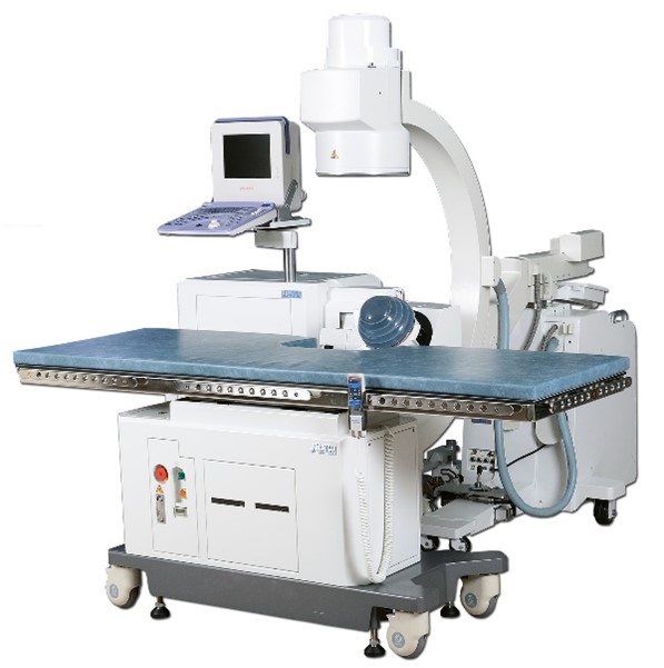
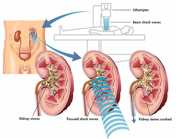
Extra Corporeal Shock Wave Lithotripsy (ESWL)
Extra Corporeal Shock Wave Lithotripsy (ESWL) is a preferred mode of treatment for kidney stones upto 1.5 cm in size. An IVU is performed before ESWL treatment to confirm an open passage from the kidney to the bladder so that finer fragments can pass out after successful treatment. The ESWL machine uses highly focused sound waves projected from outside the body to break kidney stones. As a result, the stone is reduced to sand-like particles that subsequently pass out in the urine. However, stones larger than 1.5 to 2 cm generally require more than one or two ESWL sessions.
The primary decision is whether to apply surgical treatment or wait. Removal of kidney stones by any methodology is necessary when there is evidence of obstruction. A Double J Stent insertion becomes mandatory in most of the cases.
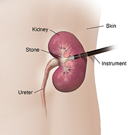
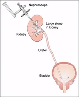
Percutaneous Nephrolithotripsy (PCNL) – Synonyms – Tunnel Surgery, Key Hole surgery for Kidney Stones
PCNL treats larger kidney stones that ESWL does not indicate for treatment. Surgeons usually perform this procedure under general, spinal, and/or epidural anesthesia. In this technique, they remove the stone by creating a small tunnel into the kidney from the back. They puncture the renal collecting system with a fine needle using X-ray and/or ultrasonography guidance and then pass a guide wire into the kidney through the needle. Next, they dilate this tract over the guide wire and insert a nephroscope (kidney telescope) into the renal pelvis. They visualize the stones, fragment them using a Swiss Lithoclast or laser, and extract them with fine forceps, which allows the kidney to become stone-free at the end of the operation in the vast majority of patients.
This is of course an operation, needing full general anesthesia, average 90 minutes of operation time, 3-4 day hospitalization, and an occasional need for blood transfusion. Patient returns to light work in 5-7 days’ time. Nevertheless, the operation is safe, for both the patients and the kidney. This operation has significantly reduced the need for open surgery (cutting surgery), which surgeons now reserve for exceptional indications.
Percutaneous Nephrolithotomy (PCNL) technique is used to treat kidney stones of:
- Large than 2.5 cms,
- Staghorn calculus,
- Calyceal diverticular calculus.