
Dr. Dilip Raja
Urologist & Andrologist
Ureteric Stone Treatment in Mumbai | Dr. Dilip Raja
Ureteric stones can cause severe pain and discomfort if not treated on time. If you’re looking for ureteric stone treatment in Mumbai, expert care can help you get quick relief and prevent complications. Advanced diagnostic methods and minimally invasive treatments ensure safe and effective stone removal.
Effective Ureteric Stone Treatment in Mumbai – Get Fast Relief from Pain Today
Get in touch with us today +91 9820074649
URETERIC STONE
The ureter is a thick-walled, narrow, cylindrical tube that connects the kidney to the urinary bladder. It measures approximately 25–30 cm in length and about 3–4 mm in diameter, and it actively transports urine from the kidney to the bladder. Additionally, the ureter has three natural narrow points where stones can become lodged, and it actively propels urine forward through rhythmic peristaltic waves.
Furthermore, ureterovesical valves prevent the backflow of urine into the kidneys, ensuring one-way movement.
Symptoms And Signs
- Incidental diagnosis on routine health check-ups.
- Dull aching pain in the back.
- Acute colic – This colicky pain begins in the flank or side of the mid-back and radiates forward to the groin (from loin to groin). It is often more severe than labor pain and is typically accompanied by nausea, vomiting, and gaseous distension.
- Urinary tract infection.
- Increased frequency of urine.
- Pain and or burning while passing urine.
- Passage of blood in urine (Haematuria).
Investigations
- CBC, Renal Profile
- Routine urine analysis
- Crystals in urine
- Blood Cells
- Puss cells in urine
- Urine for Culture Sensitivity – To rule out UTI and select best antibiotics to treat the infection.
- Ultrasonography of Kidney, Ureter and Bladder – To show the size and swelling (Hydronephrosis) of the kidney and the ureter in obstructive uropathy. It does not give information of function of kidney.
- Intravenous Urography (IVU) – To detect the size and site of stone along with function of kidney. This is a specialized test in which doctors take a series of X-rays after they inject a special contrast dye with radiopaque properties. As the kidneys excrete the contrast, it outlines them clearly on the X-rays, and clinicians capture serial images to assess their structure and function.
This is a very useful test. It gives lot of information including size and shape of kidneys, function of kidney – comparative and individual, presence of obstructive uropathy, delineates the anatomy of kidney, ureter & bladder etc. - CT Scan of Abdomen Pelvis /CT Urography – with or without oral or intravenous contrast. It also gives density of the renal stones in Hounsfield units ( HU). It also gives the function of the kidney. CT scan without contrast has some limitations.
Treatment Of Ureteric Stones
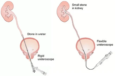
Ureterorenoscope (URS)
Ureteroscopy is highly successful procedure for the retrieval of stone in the ureter. Urologists routinely perform this procedure by passing an instrument through the natural urinary opening, into the bladder, and then into the ureter. Doctors most commonly use this procedure to treat ureteric calculi; therefore, they recommend it when ESWL or conservative treatment fails to manage the condition effectively.
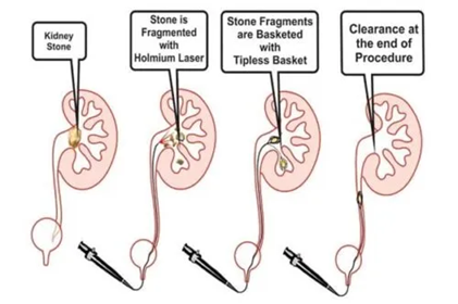
Ureterorenoscopy (URS) involves the passage of an instrument namely Ureteroscope through normal urinary passage. Under anaesthesia, the surgeon advances a ureterorenoscope through the natural urinary passage under direct vision and guides it up the ureter to the site of the stone. Once the stone is located, the surgeon chooses the appropriate treatment method. If the stone is small, they grasp it with forceps and remove it; however, if it is larger, they break it into tiny fragments using laser technology. Finally, they usually place a Double J stent after the procedure to help drain the kidney and ensure smooth urine flow.. It is a very safe procedure in experienced hands and Ureterorenscopy can treat almost all the ureteric stones.
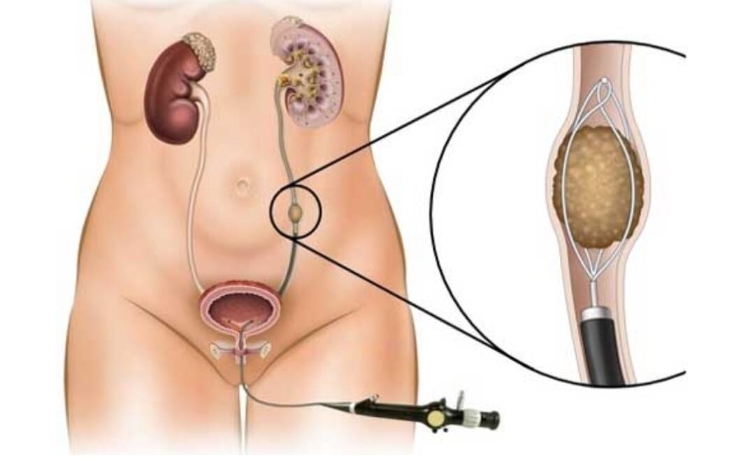
Doctors often use URS as a diagnostic tool to detect stones and, in addition, to evaluate ureteric cancer (tumors). It is a minimally invasive method of treating kidney and ureteric stone.
Ureteroscopy are of two types:
- Rigid Ureteroscopy
- Flexible Ureteroscopy
Retrograde Intrarenal Ureteroscopic Surgery (RIRS)
Doctors now perform flexible ureterorenoscopy; moreover, they pass a flexible ureteroscope through the normal ureter opening all the way up to the kidneys. As a result, they fragment stones in the renal calyces into fine particles using laser technology. This procedure requires general, spinal, or epidural anaesthesia; therefore, the anaesthesia team administers appropriate support. Finally, patients usually stay in the hospital for only 1 to 2 days.
Open Surgery
Open Surgery for Ureteric Stone are extremely rare nowadays. However, doctors may require them for large ureteric calculi that are not amenable to any endoscopic procedure.